Representations & Colors
Build a clear molecular scene by choosing how each component is drawn and colored.
A representation controls geometry: ribbon, atoms, bonds, points, or a surface. A color scheme controls meaning: element, chain, residue, secondary structure, sequence, hydrophobicity, or a uniform color. Set both per component so the visual hierarchy matches the scientific question.
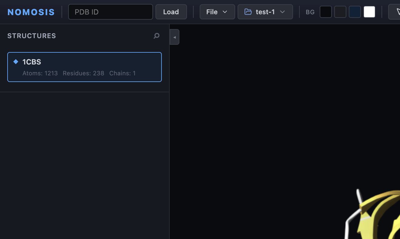
A readable protein–ligand overview: protein as Cartoon / Ribbon colored by secondary structure, ligand as Spacefill (CPK) colored by element, and water hidden.
Representation and color answer different questions
First choose how the component is drawn, then choose what its colors mean. Changing one does not automatically change the other.
Start with the question
| Scientific question | Recommended representation | Recommended color |
|---|---|---|
| What is the overall protein fold? | Cartoon / Ribbon | Secondary Structure or Chain |
| Which atoms form a ligand contact? | Protein Ribbon with atom overlay; ligand Ball & Stick | Protein Uniform; ligand Element |
| How tightly is a ligand packed? | Protein Surface or Ribbon; ligand Spacefill (CPK) | Contrasting uniform protein and element-colored ligand |
| How do chains differ? | Cartoon / Ribbon | Chain |
| Where are hydrophobic regions? | Surface | Hydrophobicity |
| How does atomic volume fill a pocket? | Spacefill (CPK) | Element or Uniform |
| How can I keep a very large system responsive? | Line, Point, or Backbone where appropriate | A simple scheme such as Chain or Uniform |
Change a component's representation
The Protein style buttons provide fast global choices for the protein: Ribbon, Atoms, Licorice, Spacefill, and Surface. Use the per-component dropdown when you need a more specific representation or are styling a ligand, water, ion, or custom element.
Representation reference
| Representation | What it emphasizes | Typical use |
|---|---|---|
| Cartoon / Ribbon | Fold and secondary structure | Proteins and nucleic acids |
| Ball & Stick | Atoms, bonds, and local geometry | Ligands, residues, measurement context |
| Licorice | Bonds with reduced atom volume | Dense atomic detail |
| Spacefill (CPK) | Van der Waals volume and packing | Steric fit and pocket occupancy |
| Surface | Smooth molecular envelope | Overall shape and pocket context |
| Mol. Surface | Detailed molecular boundary | Accessibility and molecular envelope inspection |
| Backbone | Simplified chain trace | Large polymers and comparisons |
| Line | Lightweight bond network | Large systems and contextual detail |
| Point | Minimal atom markers | Very large or dense systems |
| Putty | Variable-width backbone trace | B-factor or flexibility-oriented views |
| Ellipsoid | Anisotropic displacement | Structures with appropriate displacement data |
Not every representation is equally meaningful for every component. If a change produces an empty or confusing view, restore a standard choice such as Cartoon / Ribbon for protein or Ball & Stick for ligand.
Change a color scheme
| Color scheme | Meaning | Good use |
|---|---|---|
| Element | Standard color by atomic element | Ligands and atom-level inspection |
| Chain | A distinct color for each chain | Oligomers and interfaces |
| Residue Name | Color by residue identity | Sequence composition and local chemistry |
| Secondary Structure | Color by helix, sheet, and other structural class | Protein fold communication |
| Hydrophobicity | Color by hydrophobic character | Binding surfaces and pockets |
| Sequence ID | Color along sequence position | Following the chain from start to end |
| Uniform | One color selected by you | Visual emphasis and controlled comparison |
Build a readable scene
A professional scene usually has one dominant subject and one or two supporting layers.
Color is most useful when it carries one consistent meaning. Avoid using several unrelated color schemes in the same scene unless you explain them explicitly.
Common scene recipes
Protein–ligand overview
- Protein: Cartoon / Ribbon, Secondary Structure or a muted Uniform color.
- Ligand: Ball & Stick, Element.
- Water: hidden initially.
- Camera: Fit → All (reset), then rotate to expose the ligand.
Binding-site review
- Protein: Ribbon with Show Atoms + Ribbon enabled.
- Ligand: Ball & Stick or Spacefill (CPK), Element.
- Nearby residues: a custom element or surroundings component in Ball & Stick.
- Interactions: enable only the types being discussed.
Chain comparison
- Protein: Cartoon / Ribbon, Chain.
- Ligands: Ball & Stick, Element.
- Hierarchy: hide chains not included in the comparison.
- Camera: use the same orientation when comparing saved scenes.
Avoid these mistakes
- Do not use a surface as the only representation if you need to discuss residue-level geometry.
- Do not hide a component and assume its data was removed; visibility is reversible.
- Do not choose an atom-level representation for an entire large simulation unless the added detail is necessary.
- Do not reuse nearly identical uniform colors for components that must be distinguished.
- Do not change the active structure accidentally when several structures are loaded.